Mark Kopec Now
MRI
What is an MRI?
Magnetic Resonance Imaging (MRI) is a diagnostic imaging technique that uses powerful magnets and radio waves to create detailed images of the body’s organs and tissues.
Unlike X-rays, MRI does not use ionizing radiation. Instead, it relies on the magnetic properties of hydrogen atoms in the body to produce images.
A Brief History of MRI
The concept of MRI arose in the 1940s, but it wasn’t until the 1970s that the first practical MRI scanner appeared. Raymond Damadian, a physician, is credited with the initial idea of using MRI for medical imaging. Paul Lauterbur and Peter Mansfield made significant contributions to the development of the technology, leading to its widespread use in clinical settings by the 1980s.

Who Orders MRIs and Why?
A variety of doctors and other medical professionals order MRIs, including:
- Radiologists: Specialists in medical imaging.
- Orthopedists: Doctors specializing in musculoskeletal conditions.
- Neurologists: Doctors specializing in the nervous system.
- Oncologists: Doctors specializing in cancer treatment.
- Cardiologists: Doctors specializing in heart conditions.
Doctors generally order MRIs to diagnose a wide range of conditions, including:
- Brain tumors
- Spinal cord injuries
- Joint damage
- Muscle tears
- Ligament injuries
- Bone fractures
- Internal bleeding
- Infections
- Cardiovascular abnormalities
The MRI Procedure
MRIs are typically administered by radiologic technologists, who are trained in positioning patients and operating the MRI machine. The procedure involves:
- Preparation: Patients are asked to remove any metal objects, as these can interfere with the magnetic field. They may also be given a contrast agent to enhance image quality.
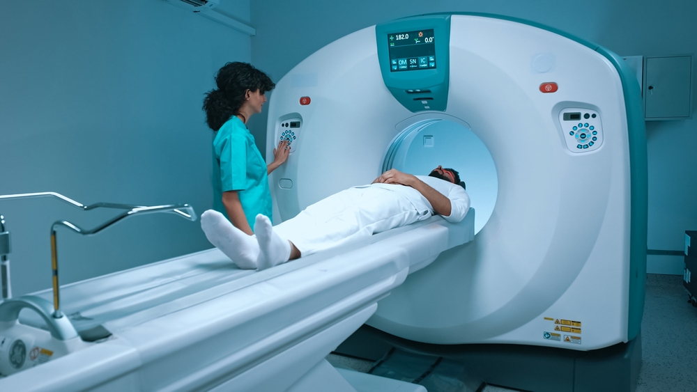
- Scanning: The patient lies on a table that slides into the MRI machine. The machine creates a strong magnetic field, and radio waves are used to produce images. The procedure is typically painless, but it can be noisy.
- Imaging: The MRI machine creates detailed images of the targeted body part. A radiologist then interprets these images.
Doctors can target MRIs to specific body parts or full-body scans, depending on the reason for the exam.
What MRI Can and Cannot Show
MRIs excel at visualizing soft tissues, such as the brain, spinal cord, muscles, and ligaments. They can provide detailed information about the structure and function of these tissues. However, MRIs may not be as effective at visualizing bones or detecting certain types of tumors.
MRIs can diagnose a wide range of conditions, but they are not infallible. False positives and false negatives can occur. Therefore, doctors should always interpret MRI results in conjunction with other diagnostic tests and clinical findings.
The MRI Report
The MRI report, prepared by a radiologist, generally includes:
- Patient information
- The reason for the exam
- A detailed description of the findings
- Impressions and also diagnoses
- Recommendations for further testing or treatment
MRIs in Medical Malpractice Cases
Medical malpractice cases involving MRIs can arise from various errors, including:
- Failure to order an MRI when indicated: This can lead to misdiagnosis or delayed diagnosis and worsened patient outcomes.
- Incorrect interpretation of MRI results: Misreading of images can result in misdiagnosis or improper treatment.
- Patient injury during the MRI procedure: This can occur due to equipment malfunction, human error, or failure to screen patients for metal implants or claustrophobia.
- Doctors fail to disclose risks associated with the MRI procedure: Patients have the right to have doctors inform them about potential complications before undergoing the exam.
If you have a potential medical malpractice case, then visit the Kopec Law Firm free consultation page or video. Then contact us at 800-604-0704 to speak directly with Attorney Mark Kopec. He is a top-rated Baltimore medical malpractice lawyer. The Kopec Law Firm is in Baltimore and pursues cases throughout Maryland and Washington, D.C.


